How does the Vi-CELL XR differentiate viable versus non-viable cells?
Service AI
What does a VI-cell do?
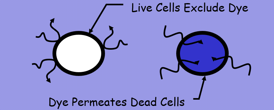
The Vi-CELL XR utilizes the widely accepted Trypan Blue Dye Exclusion Method, in which the analysis relies upon the fact that as cells die and become non-viable, their membranes become permeable. The Trypan Blue dye molecules when mixed with the cells will enter the dead cells, but cannot penetrate the viable or live cells as their membranes remain intact. As a result of which when the dead cells are viewed using the Vi-CELL or a microscope are stained and therefore considerably darker than the viable/live cells. The analysis uses the difference in color or darkness to differentiate between live and dead cells.
The image capture device in the Vi-CELL XR is a CCD (Charged Coupled Device) housed in an excellent quality fire wire video camera with associated optics allows the user to view the cells. The CCD device consisting of 1392 x 1040 CCD array comprises of a large number of pixels, which are individual light detectors, that record the amount of light that they are exposed to. Each detector will record a different amount of light. The different amount is measured on what is known as a "gray scale".
If you are interested in purchasing a fully re-certified Beckman Vi-CELL XR that we currently have in our inventory, contact us at support@serviceai.us.
Please do not hesitate to ask us what it means to re-certify an instrument, and the steps we will be taking to recertify the Beckman Vi-CELL XR before placing it in your laboratory.
Vi-CELL XR
Automated cell viability analyzers such as the Beckman Coulter Vi-CELL XR offer several advantages over manual cell counting.


